
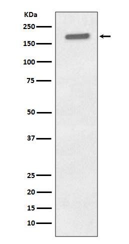
Western blot analysis of Phospho-EGFR (Y1197) expression in A431 cell lysate treated with EGF.
all(4)
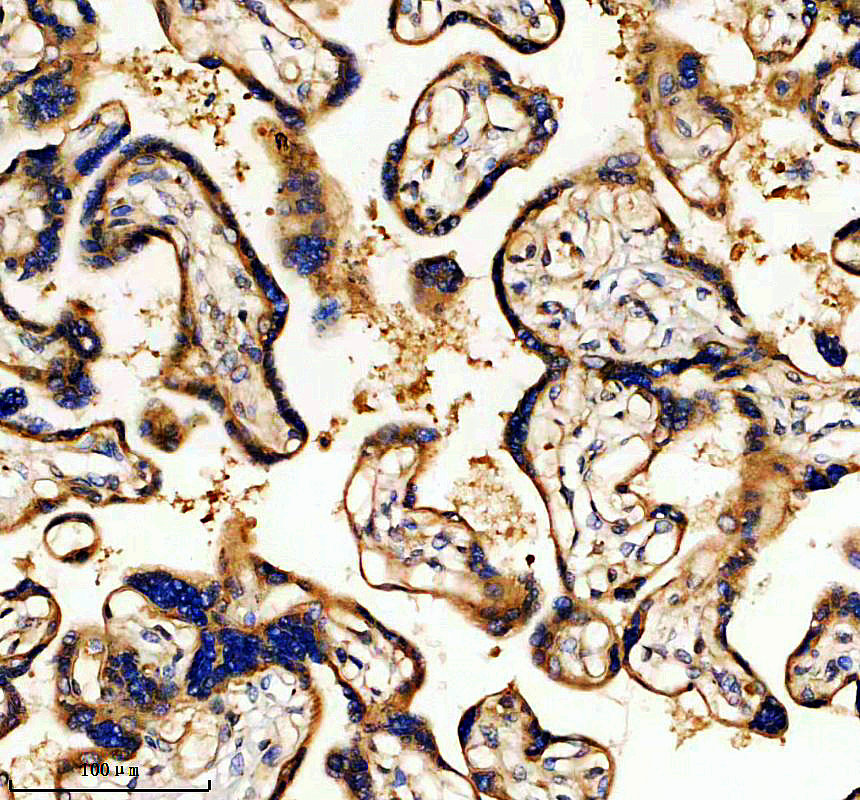
IHC analysis of EGFR using anti-EGFR antibody (BM4453).
EGFR was detected in a paraffin-embedded section of human placenta tissue. The tissue section was incubated with rabbit anti-EGFR Antibody (BM4453) at a dilution of 1:200 and developed using HRP Conjugated Rabbit IgG Super Vision Assay Kit (Catalog # SV0002) with DAB (Catalog # AR1027) as the chromogen.
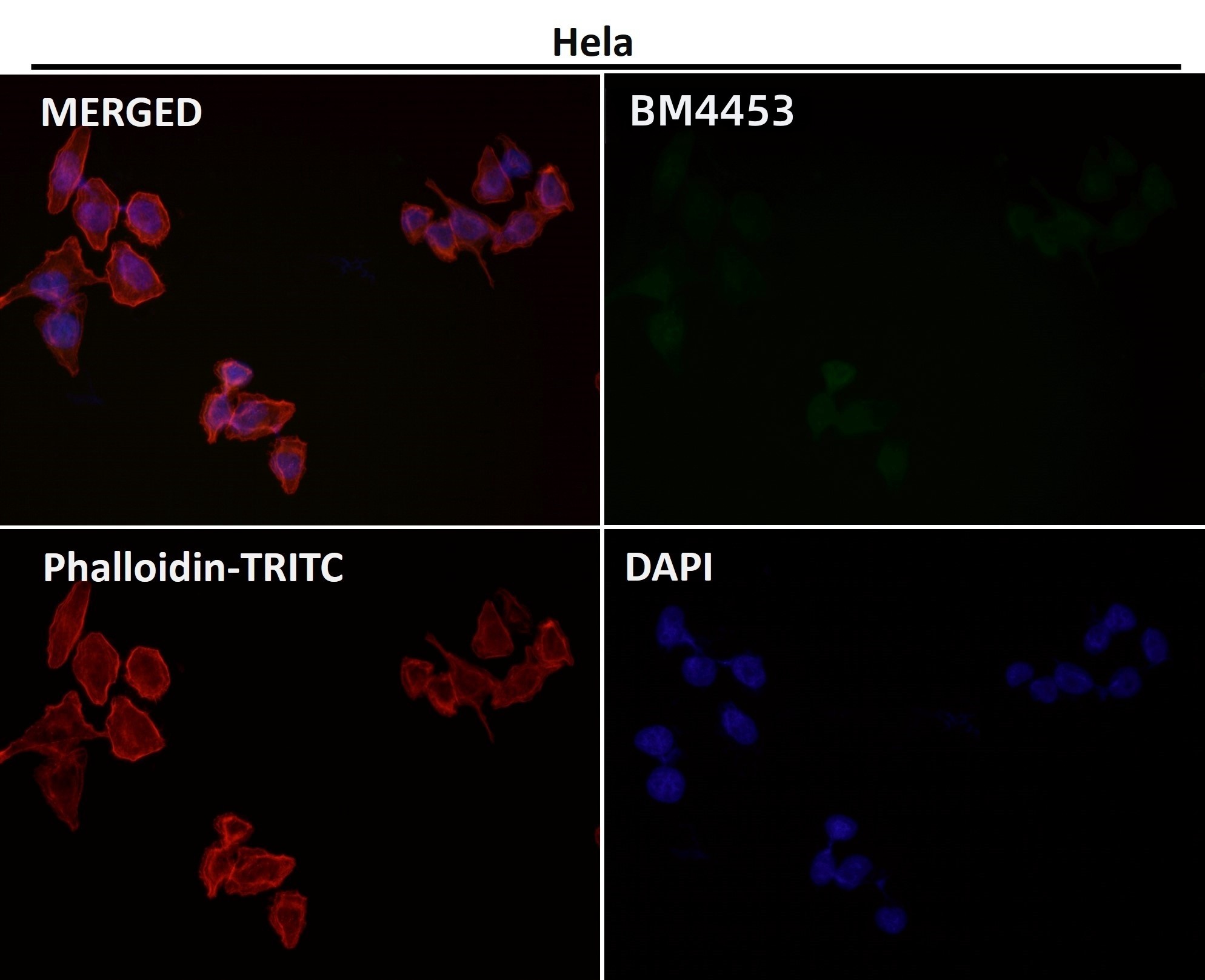
Immunofluorescent analysis using the Antibody.
all(4) 
| Western blot (WB): | 1:500-2000 |
| Immunohistochemistry (IHC): | 1:50-200 |
| Immunocytochemistry/Immunofluorescence (ICC/IF): | 1:50-200 |
| ImmunoPrecipitation (IP): | 1:20 |
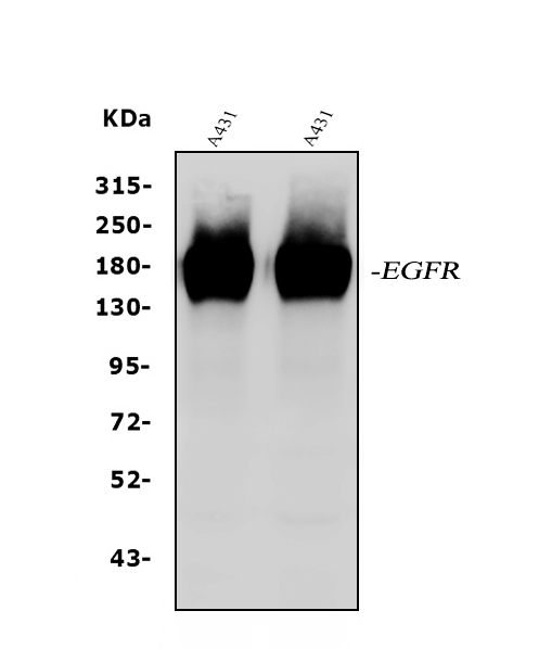
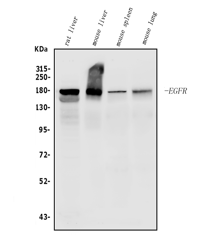
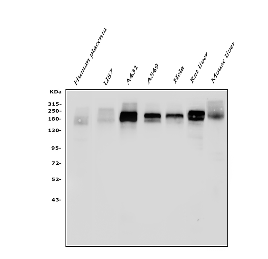
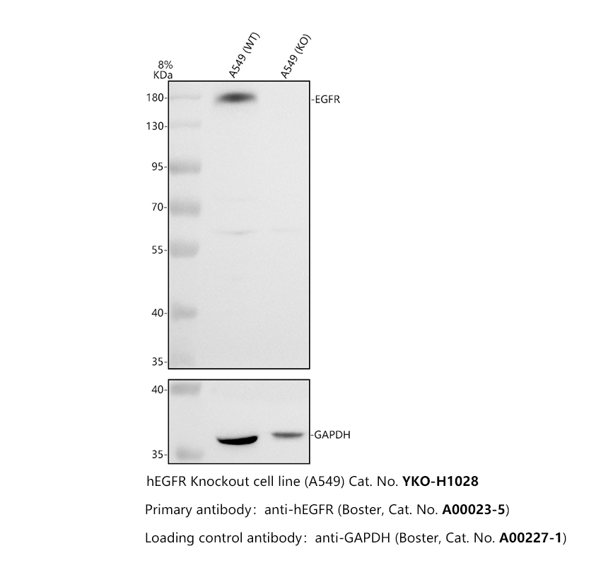
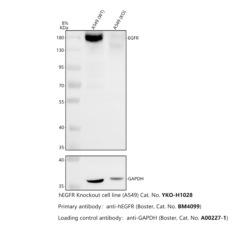
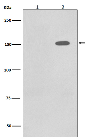
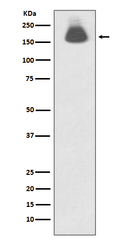
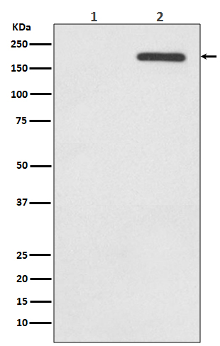
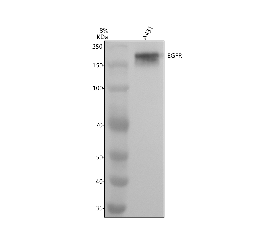
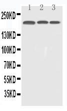
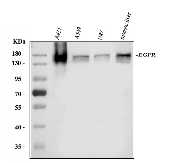
Western blot analysis of Phospho-EGFR (Y1197) expression in A431 cell lysate treated with EGF.
IHC analysis of EGFR using anti-EGFR antibody (BM4453).
EGFR was detected in a paraffin-embedded section of human placenta tissue. The tissue section was incubated with rabbit anti-EGFR Antibody (BM4453) at a dilution of 1:200 and developed using HRP Conjugated Rabbit IgG Super Vision Assay Kit (Catalog # SV0002) with DAB (Catalog # AR1027) as the chromogen.
Immunofluorescent analysis using the Antibody.
Western blot analysis of Phospho-EGFR (Y1197) expression in A431 cell lysate treated with EGF.
IHC analysis of EGFR using anti-EGFR antibody (BM4453).
EGFR was detected in a paraffin-embedded section of human placenta tissue. The tissue section was incubated with rabbit anti-EGFR Antibody (BM4453) at a dilution of 1:200 and developed using HRP Conjugated Rabbit IgG Super Vision Assay Kit (Catalog # SV0002) with DAB (Catalog # AR1027) as the chromogen.
Immunofluorescent analysis using the Antibody.
 联系我们
联系我们027-67845390
 关注我们
关注我们
本司产品仅用于科研,不用于临床诊断和治疗
联系方式:027-67845390/1/2 技术支持:武汉丰网
© 1993-2026 Boster Biological Technology co.Itd E-mail:boster@boster.com
鄂ICP备05005548号-2
 鄂公网安备 42018502007312号

 积分商城
积分商城  购物车
购物车  登录/注册
登录/注册  您当前的位置:
  说明书
说明书 一键复制产品信息
 成功添加到购物车
 微信客服
微信客服


 微信扫一扫立即咨询
